
At a Glance
- Post-liposuction and body contouring recovery involves mechanical disruption of superficial lymphatic channels, persistent protein-rich interstitial edema, seroma risks, and dense subcutaneous fibrosis bands that compromise cosmetic outcomes if untreated.
- Inner ball roller compressive microvibration provides gentle, non-thermal oscillating pressure cycles (40–150 Hz) and rotational shear forces that stimulate lymphatic transport, resolve indurated fibrosis, and promote skin retraction without traction or epidermal thermal risks.
- Clinical protocols follow a three-stage postoperative timeline: Stage 1 (Weeks 2–3, ultra-low pressure lymphatic drainage), Stage 2 (Weeks 4–6, moderate shear for active fibrosis softening), and Stage 3 (Week 8 onward, contour refinement and tissue tightening).
- Unlike manual lymphatic drainage (MLD), which imposes severe physical strain on therapists and high session-to-session variability, inner ball roller machines standardize treatment depth and pressure via digital sensor feedback while accelerating patient volume recovery.
- The multi-applicator configuration—combining small (60–80mm), medium (100–120mm), and large (140–180mm) roller assemblies with real-time pressure monitoring—ensures customized anatomical coverage across the abdomen, flanks, back, and extremities.
- MNLT Laser manufactures clinical-grade inner ball roller systems compliant with CE, FDA, EU MDR, and ISO 13485 standards, operating in over 20,000 surgical rehabilitation and aesthetic centers across 120 countries.
Surgical body contouring procedures—most notably tumescent liposuction, high-definition lipo-sculpture, abdominoplasty (tummy tuck), and brachioplasty—have surged globally, representing some of the most frequently performed cosmetic surgeries. However, plastic surgeons and aesthetic clinic directors recognize that the surgical theater accounts for only half of the aesthetic outcome; the subsequent postoperative recovery phase dictates whether a patient achieves smooth, defined contours or suffers from chronic edema, contour irregularities, seroma formation, and disfiguring subcutaneous fibrosis. While manual lymphatic drainage (MLD) has historically served as the post-surgical aftercare benchmark, the sheer physical exhaustion of repetitive manual massage and significant inter-practitioner variability limit clinical scalability. Inner ball roller compressive microvibration has rapidly established itself as the modern clinical standard for post-surgical physical rehabilitation. This comprehensive clinical guide examines the pathophysiology of post-cannulation tissue trauma, delineates timeline-based treatment protocols, details anatomical applicator selection, and presents comparative efficacy frameworks for postoperative clinics in 2026.
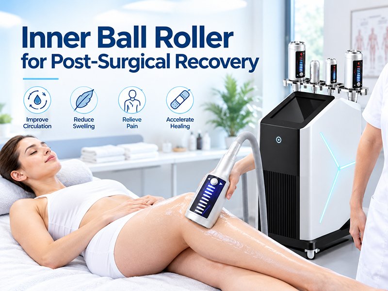
Pathophysiology of Surgical Tissue Trauma: The Post-Liposuction Milieu
To safely deploy mechanical rehabilitation modalities, practitioners must understand the biological microenvironment left behind by surgical cannulas. Liposuction is an inherently traumatic mechanical event within the subcutaneous hypodermis.
1. Vascular & Lymphatic Disruption
During suction-assisted or energy-based (VASER, laser) liposuction, rapid back-and-forth cannula excursions avulse fat lobules while shearing delicate terminal blood capillaries and initial lymphatic collectors. The severed lymphatic network cannot efficiently evacuate the influx of interstitial fluid, surgical tumescent fluid remnants, and extravasated red blood cells. This results in intense, brawny postoperative edema that pools in dependent anatomical spaces.
2. Protein-Rich Stasis and Inflammatory Fibrin Deposition
Stagnant interstitial fluid in post-surgical beds is rich in cellular debris, inflammatory cytokines, and plasma proteins. If this fluid remains static for prolonged periods, it precipitates a persistent low-grade inflammatory state. Macrophages, neutrophils, and activated fibroblasts infiltrate the interstitial matrix, depositing dense disorganized collagen type I and III fibers. This process leads directly to subcutaneous fibrosis—manifesting clinically as hard, painful lumps, sheet-like tissue induration, and surface rippling that distort the final surgical silhouette.
3. Fascial Adhesions and Skin Retraction Deficits
Following the evacuation of deep and superficial fat layers, the overlying cutaneous flap must readapt and contract against the underlying deep muscular fascia. In areas where fibrous adhesions form irregularly, the skin adheres prematurely in tethered patches, creating visible divots, asymmetry, and skin laxity.
For a foundational understanding of how compressive microvibration interacts with chronic fluid stagnation and deep interstitial channels, review our clinical analysis of inner ball roller for lymphedema management.
Biological Action of Compressive Microvibration in Surgical Tissue Repair
Compressive microvibration exerts specific physical forces that counteract post-surgical complications through non-thermal, non-suction biomechanical vectors:
- Hydraulic Mobilization Without Suction: Traditional vacuum-assisted endermology machines apply negative pressure (suction) to lift tissue. In early post-surgical states, negative pressure traction risks tearing fragile neo-vascular beds, increasing bruising, and worsening seroma pockets. Compressive microvibration utilizes positive rhythmic compression, gently pumping fluid forward into functional collateral lymphatic basins without pulling on healing surgical planes.
- Mechanical Breakdown of Early Fibrotic Cross-Links: The 360-degree rotating cylinder generates continuous tangential shear forces in the superficial hypodermis. This multidirectional micromassage mechanically breaks down nascent fibrin deposits and disorganized collagen bundles before they can calcify into permanent fibrotic sheets.
- Vascular Re-perfusion and Neocollagenesis: Low-frequency rhythmic oscillations stimulate endothelial nitric oxide (eNOS) release, inducing gentle physiological vasodilation that enhances local tissue oxygenation and metabolic clearance, expediting skin flap re-adherence.
Practitioners seeking a detailed engineering review of rotation parameters, frequency ranges, and microvibration kinetics should consult our inner ball roller compressive microvibration technical guide.
Post-Surgical Timeline Protocols: When and How to Treat
The most critical clinical rule in post-surgical rehabilitation is respecting tissue healing phases. Applying aggressive pressure too early risks reopening surgical channels, while intervening too late allows mature fibrosis to set permanently.
| Rehabilitation Phase | Timeline Window | Primary Clinical Objective | Applicator & Pressure Setting | Session Frequency |
|---|---|---|---|---|
| Stage 1: Acute Drainage | Weeks 2–3 (Post-Op Day 14+) | Gentle interstitial fluid evacuation, seroma prevention, hematoma resolution | Medium/Large roller; ultra-light pressure (10–15 mmHg); low speed (40–60 Hz) | 2–3 sessions per week; 20–30 min per zone |
| Stage 2: Anti-Fibrosis | Weeks 4–7 | Breaking active subcutaneous fibrosis bands, softening tissue lumps, fascial release | Medium roller; moderate pressure (20–30 mmHg); medium speed (70–100 Hz) | 2 sessions per week; 30–40 min per zone |
| Stage 3: Remodeling & Contouring | Week 8+ onward | Skin retraction, dermal tightening, contour smoothing, neuromuscular toning | Full suite (Large/Medium + EMS handle); moderate-high pressure; high speed (100–150 Hz) | 1–2 sessions per week; 40 min per zone |
Phase 1: Acute Decongestive Phase (Weeks 2–3)
- Entry Criteria: Surgical port sites and incisions must be fully closed and re-epithelialized with no active seroma discharge or signs of infection. Written consent from the operating plastic surgeon is mandatory.
- Technique: Extremely gentle gliding along anatomical lymphatic vectors toward intact regional nodal hubs (axillary or inguinal). The operator must never apply downward manual force; the intrinsic weight of the handpiece provides sufficient displacement. Avoid direct manipulation over healing scar lines.
Phase 2: Active Fibrotic Remodeling Phase (Weeks 4–7)
- Clinical Focus: This is the critical therapeutic window. Fibroblastic activity peaks during this period. Practitioners identify palpable subcutaneous nodules (fibrosis cords) and systematically work the roller assembly in cross-hatch, multidirectional strokes.
- Technique: Controlled, rhythmic pressure mobilizes dense collagen matrix clusters, breaking micro-adhesions without tearing connective tissue.
Phase 3: Skin Retraction & Muscular Consolidation (Week 8 Onward)
- Clinical Focus: Addressing residual cutaneous laxity and refining muscular outlines. In modern clinical suites equipped with an EMS handle, practitioners can transition from passive mechanical drainage to active neuromuscular stimulation.
- Technique: High-frequency rotation coupled with low-frequency electrical muscle stimulation contracts the underlying deep muscular bed while simultaneously firming dermal tissue, ensuring tight adherence between the cutaneous flap and muscular wall.
Compressive Microvibration vs. Traditional Post-Surgical Modalities
Surgical aftercare centers typically utilize a mix of manual therapies, radiofrequency, and ultrasound. Understanding the competitive advantages of compressive microvibration assists medical directors in streamlining equipment investments.
| Feature / Clinical Outcome | Inner Ball Roller System | Manual Lymphatic Drainage (MLD) | External Therapeutic Ultrasound | Negative-Pressure Vacuum Roller |
|---|---|---|---|---|
| Physical Mechanism | Positive compressive microvibration + 360° rotation | Manual skin stretching along lymphatic channels | Acoustic sound wave micro-cavitation / thermal | Negative pressure suction + motorized rollers |
| Risk in Early Recovery (Weeks 2–3) | Very low (non-thermal, zero skin traction) | Extremely low (gold standard manual safety) | Moderate (risk of deep internal thermal injury) | High (traction can tear neo-vessels & cause seroma) |
| Efficacy Against Dense Fibrosis | Exceptional (rotational shear physically softens bands) | Moderate to low (requires intense deep manual pressure) | Moderate (softens tissue via acoustic energy) | Moderate (can cause bruising on fibrotic tissue) |
| Practitioner Physical Fatigue | Very low (machine provides all kinetic motion) | Extremely high (limits daily therapist capacity to 3–4 clients) | Low | Low to moderate |
| Treatment Standardization | High (reproducible digital frequency and sensor pressure) | Low (highly dependent on individual manual technique) | High (timer and power settings) | Moderate |
| Skin Tightening Stimulation | High (microvibration stimulates collagen synthesis) | None (pure fluid drainage) | Low | Moderate |
For clinics treating a broad demographic that includes both aesthetic cellulite clients and surgical recovery patients, compare these protocols against our inner ball roller cellulite treatment clinic guide.
Anatomical Protocol Design: Abdomen, 360-Lipo, and Extremities
Different surgical harvest sites present distinct anatomical hurdles:
1. The Abdomen & Flanks (Lipo 360 + Tummy Tuck)
- Anatomical Hurdle: In patients who underwent concurrent abdominoplasty, the inferior lymphatic drainage pathways leading to the inguinal nodes are severed across the lower abdominal incision. All abdominal lymphatic fluid must be rerouted retrograde or superiorly toward the axillary basins and parasternal nodes.
- Technique: The operator uses the large roller assembly (140–180mm), moving in upward, sweeping strokes from the suprapubic zone toward the bilateral axillary basins. For lateral flanks, oblique strokes channel fluid along the lateral trunk toward the scapular and axillary nodes.
2. Inner & Outer Thighs
- Anatomical Hurdle: The medial thigh skin is remarkably thin and prone to post-surgical skin waviness, while the lateral thigh (trochanteric area) is a prime location for stubborn fibrotic plateaus.
- Technique: Use the medium roller (100–120mm) on the outer thigh with cross-hatched strokes to soften fibrosis. On the inner thigh, utilize light sweeping strokes directed upward toward the inguinal crease to relieve chronic dependent fluid collection around the knee.
3. Arms (Brachioplasty or Arm Lipo)
- Anatomical Hurdle: Prone to persistent dependent edema extending into the forearm and wrist.
- Technique: Deploy the small roller applicator (60–80mm), beginning distal to the elbow to drain pooled fluid, progressing proximal-superiorly toward the axilla.
Clinical Safety Boundaries & Absolute Contraindications
While compressive microvibration is non-invasive, surgical aftercare requires strict adherence to clinical safety boundaries:
Absolute Contraindications
- Unhealed Incisions or Open Drainage Ports: Treatment must never pass directly over active incisions, scabs, or open drain insertion sites until complete primary wound closure is verified.
- Suspected Deep Vein Thrombosis (DVT): Surgical patients (especially post-abdominoplasty and thigh liposuction) carry heightened DVT risks. Any unilateral asymmetric swelling accompanied by calf tenderness, warmth, or erythema demands immediate cessation of treatment and ultrasound evaluation.
- Active Infection or Cellulitis: Presence of localized fever, spreading erythema, or purulent exudate requires prompt surgical referral and antibiotic therapy.
- Active, Unaspirated Seroma: If a fluid collection is fluctuant upon palpation, mechanical rolling will simply displace the fluid pocket without resolution and may prevent pocket adhesion. The operating surgeon must aspirate the seroma before compressive therapy commences.
Best Practice Quality Control
- Pre-Treatment Photography & Circumferential Measurement: Document baseline tissue density, bruising extent, and limb circumference at identical anatomical landmarks prior to every session.
- Compression Garment Discipline: Patients must re-don their medical-grade post-surgical compression garments (Faja Stage 1 or 2) immediately following every compressive microvibration session to prevent interstitial fluid re-accumulation.
Why Surgical Rehabilitation Centers Choose MNLT Inner Ball Roller
Engineering integrity dictates clinical reliability. MNLT Laser (Shandong Moonlight Electronic Technology Co., Ltd.) brings over 19 years of medical device manufacturing excellence to surgical recovery centers worldwide:
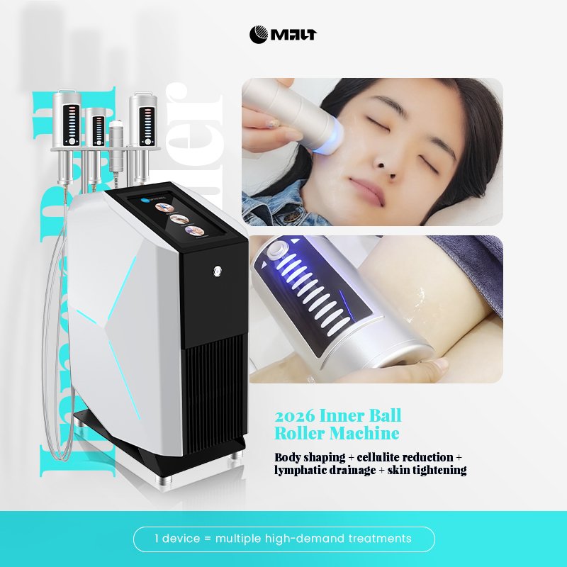
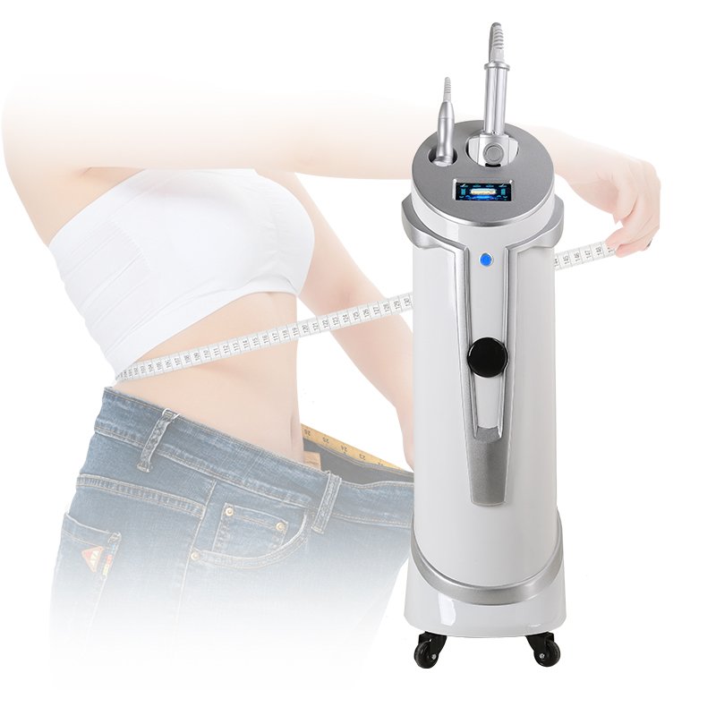
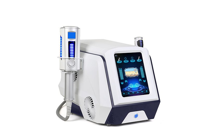
- Triple-Applicator Versatility: Each clinical platform includes small, medium, and large roller assemblies, ensuring seamless transitions between delicate cervical/brachial areas and broad abdominal zones.
- Integrated Dynamic Pressure Sensors: Real-time sensor feedback provides practitioners with continuous visual readouts of applied downward pressure, ensuring uniform, non-traumatic treatments across multiple clinic staff members.
- Modular Multi-Frequency Control: Precise electronic regulation from 40 Hz to 150 Hz allows smooth progression from delicate early-stage lymphatic drainage to aggressive late-stage fibrotic remodeling.
- Comprehensive Global Certifications: Full certification under CE, US FDA, EU MDR, and ISO 13485 ensures complete compliance with hospital audit requirements and professional insurance standards.
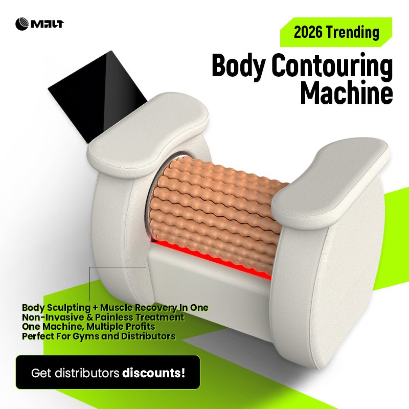
Explore our full clinical portfolio on the inner ball roller machine product category page, or contact MNLT’s medical engineering team today for specialized technical specifications and direct manufacturing procurement.
Inquire Now
Fill out the form for more information, and an MNLT product manager will contact you shortly!
Frequently Asked Questions
When can a patient safely start inner ball roller treatments after liposuction?
Patients can typically begin Stage 1 treatments between post-operative days 14 and 21 (Weeks 2–3), provided that all surgical incisions and drain entry sites are fully healed, closed, and free from active drainage or infection. Written medical clearance from the operating plastic surgeon is always recommended prior to initiating therapy. During this early phase, treatments must use the lowest frequency and ultra-light pressure focused purely on gentle lymphatic evacuation.
How does inner ball roller resolve subcutaneous fibrosis and lumps after surgery?
Subcutaneous fibrosis develops when static, protein-rich post-surgical fluid triggers fibroblasts to deposit dense, disorganized collagen fibers, forming hard palpable lumps beneath the skin. Inner ball roller technology resolves this through non-thermal compressive microvibration combined with 360-degree rotational shear forces. The continuous mechanical displacement gently breaks down nascent collagen cross-links, softens hardened tissue bands, and restores normal tissue pliability without risking dermal thermal injury.
Why is compressive microvibration preferred over negative-pressure vacuum massage post-surgery?
Negative-pressure vacuum devices rely on suction to lift and stretch the tissue. In post-surgical patients, applying tensile traction to freshly undermined skin flaps can tear newly forming microvascular beds, provoke secondary bleeding, worsen bruising, and significantly increase the risk of seroma pocket formation. Inner ball roller uses positive, pulsed compression and rhythmic rolling that moves fluid forward into lymphatic pathways without pulling or separating healing anatomical tissue planes.
How many post-surgical sessions are typically required for optimal recovery?
A standard post-liposuction rehabilitation protocol consists of 10 to 15 sessions. The typical schedule begins with 2 to 3 sessions per week during Weeks 2 through 5 to manage acute edema and prevent early fibrosis formation. Frequency then tapers to 1 to 2 sessions per week through Weeks 6 to 10 for deep tissue remodeling, skin retraction, and final contour smoothing. Severe cases with established dense fibrosis may benefit from an extended 20-session rehabilitation program.
Can inner ball roller cause or worsen a seroma?
When used correctly according to clinical protocols, inner ball roller helps prevent seromas by continuously evacuating interstitial fluid. However, if a fluctuant, encapsulating seroma pocket has already formed, mechanical rolling over the active fluid pocket is contraindicated. Rolling over an un-aspirated seroma will not dissolve the fluid collection and may prevent the dead space from adhering. The surgeon must aspirate the fluid collection first, after which gentle compression around the perimeter can resume.
Should patients wear compression garments (fajas) after their treatment?
Yes, absolutely. The post-operative compression garment (Faja Stage 1 or 2) must be put back on immediately following each inner ball roller session. The mechanical treatment mobilizes large volumes of stagnant interstitial fluid into the lymphatic system; without immediate external counter-pressure from the medical garment, fluid will swiftly redistribute back into the treated dependent spaces, diminishing clinical results.
What regulatory certifications are necessary for post-surgical rehabilitation equipment?
Post-surgical rehabilitation clinics should demand CE marking, US FDA 510(k) clearance, EU MDR compliance, and ISO 13485 medical manufacturing certification. Because treatments are delivered to recovering post-surgical patients, verifying authentic, traceable registration numbers within public medical device registries ensures device safety, electrical shielding, and full professional indemnity compliance.
Reviewed by David Ma
CTO & Chief Engineer, MNLT Laser (Shandong Moonlight Electronics Tech Co., Ltd.)
19 years in aesthetic device manufacturing. David leads MNLT's R&D team across the full professional aesthetic device portfolio, ensuring all technical content meets the highest standards of accuracy and clinical safety.